This is a technical companion page to the main article — a full raw-data appendix containing the input clinical panels and verbatim outputs from both services across each of the five cases. Nothing is paraphrased and nothing has been tidied up. If you want to verify the experiment or run the same panels through a different LLM, the material is all here.
How to Read This Page
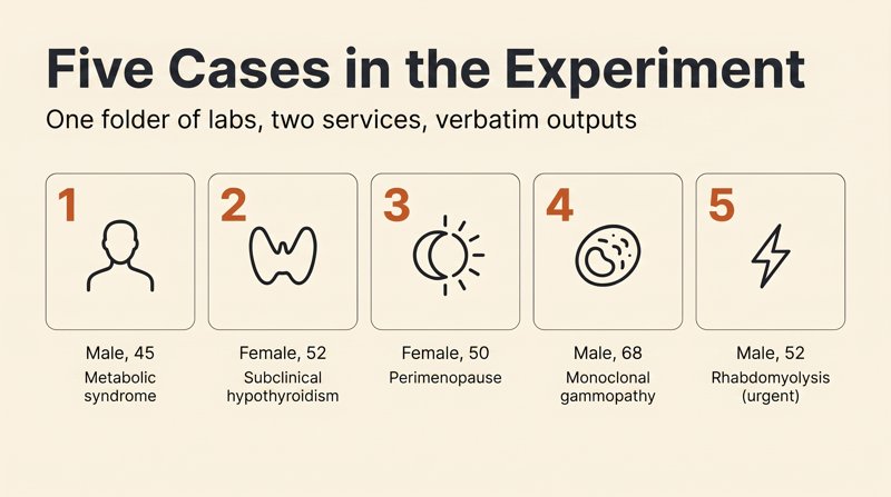
Each case has three blocks:
- Input Data — the clinical panel plus patient history loaded into both services. Reconstructed from publications in peer-reviewed journals (PubMed, Blood, Annals of Family Medicine, and others) with every clinically meaningful abnormality preserved.
- ChatGPT Response — the verbatim output from ChatGPT, Plus tier, GPT-5.4. The prompt was the same in every case: “please interpret my labs.” No follow-up, no editing.
- Wizey Response — the verbatim output from Wizey. Identical prompt.
All outputs are shown inside monospaced code blocks so it’s visible that this is raw text copied from the services’ interfaces as-is (with emojis, markers, and the original response structure).
Methodology
- All runs were performed on April 17, 2026, in a single day.
- ChatGPT — the chat.openai.com web interface, ChatGPT Plus tier, model GPT-5.4.
- Wizey — wizey.one, production version of the service.
- The prompt in both cases was identical — “please interpret my labs.”
- Panels were loaded as text (not screenshots) to eliminate OCR differences.
- Outputs were not edited and are preserved verbatim with original formatting.
- Four cases are routine outpatient scenarios; one (case 5) is urgent, included specifically to test triage behavior.
Limitations
Five cases is a pattern illustration, not a statistic. We saw the same shape in each of the five, but this is not a randomized study over a thousand runs. If someone else repeats the same runs and gets a different result, we’ll publish it. That’s how science works.
We’re the Wizey team — the conflict of interest is obvious. That’s exactly why:
- The methodology was fixed before the runs.
- All outputs are quoted verbatim — including the case where ChatGPT beat us (case 2, MGUS).
- The panels are reproducible — reconstructed from public sources.
Case 1 — 45-Year-Old Male, Metabolic Syndrome
Input Data
Patient: male, 45 years old
Occupation: marketing director
Height 180 cm, weight 94 kg, BMI 29.0
Waist circumference: 104 cm

Family history:
— Father: type 2 diabetes since age 58
— Mother: hypertension since age 55

Complaints:
— Persistent fatigue for 1–1.5 years, "wants to sleep even after weekends"
— Hard to get up in the morning despite 7 hours of sleep
— Weight gain of 6 kg over 2 years with no obvious change in diet
— Decreased libido over the past year
— Morning erections have become rare
— Episodes of irritability, loss of motivation
— Knee pain after prolonged walking
— Snoring (per wife), morning headache sometimes
— Heartburn 2–3 times a week in the evening

Lifestyle:
— Sedentary work, 10–11 hours at the computer
— Minimal physical activity — dog walks
— 1–2 glasses of red wine almost every evening "to unwind"
— Occasional spirits on weekends with friends
— Sleep 6–7 hours, often goes to bed after midnight
— High stress — running a major project
— Nonsmoker
— Coffee 3–4 cups a day

Prior workups: routine health screening 6 years ago — unremarkable. No follow-up since.

Current medications: none.

Office blood pressure: 135/88 mmHg.

═══════════════════════════════════════
COMPLETE BLOOD COUNT
═══════════════════════════════════════
Hemoglobin: 152 g/L (ref 130–170)
Red blood cells: 5.0 ×10¹²/L (ref 4.3–5.7)
Hematocrit: 44.8% (ref 38–49)
MCV: 89 fL (ref 80–100)
MCH: 30.4 pg (ref 27–33)
MCHC: 340 g/L (ref 320–360)
RDW: 13.0% (ref 11.5–14.5)
Platelets: 262 ×10⁹/L (ref 150–400)
White blood cells: 6.8 ×10⁹/L (ref 4.0–10.0)
Neutrophils: 58% (ref 45–70)
Lymphocytes: 32% (ref 19–37)
Monocytes: 7% (ref 3–11)
Eosinophils: 2% (ref 1–5)
Basophils: 1% (ref 0–1)
ESR: 8 mm/hr (ref up to 15)

═══════════════════════════════════════
CARBOHYDRATE METABOLISM
═══════════════════════════════════════
Fasting glucose: 5.9 mmol/L (ref 3.9–6.1)
HbA1c: 5.9% (ref <5.7; prediabetes 5.7–6.4)
Fasting insulin: 19 μIU/mL (ref 2.6–24.9)
C-peptide: 3.8 ng/mL (ref 1.1–4.4)
HOMA-IR: 4.9 (ref <2.5)

═══════════════════════════════════════
LIVER MARKERS
═══════════════════════════════════════
ALT: 58 U/L (ref <45)
AST: 42 U/L (ref <40)
ALP: 82 U/L (ref 40–130)
GGT: 78 U/L (ref <55)
Total bilirubin: 16 μmol/L (ref 3.4–20.5)
Direct bilirubin: 3.8 μmol/L (ref <5.1)
Albumin: 44 g/L (ref 35–52)
Total protein: 74 g/L (ref 64–83)

═══════════════════════════════════════
RENAL FUNCTION
═══════════════════════════════════════
Creatinine: 95 μmol/L (ref 62–115)
Urea: 5.2 mmol/L (ref 2.5–7.1)
eGFR (CKD-EPI): 91 mL/min/1.73 m²
Uric acid: 468 μmol/L (ref 208–428)

═══════════════════════════════════════
LIPID PROFILE (extended)
═══════════════════════════════════════
Total cholesterol: 5.8 mmol/L (ref <5.2)
LDL: 3.6 mmol/L (target <3.0)
HDL: 0.95 mmol/L (ref >1.0)
Triglycerides: 2.4 mmol/L (ref <1.7)
Non-HDL cholesterol: 4.85 mmol/L
Apolipoprotein A1: 1.24 g/L (ref 1.04–2.02)
Apolipoprotein B: 1.35 g/L (ref 0.55–1.30)
Atherogenic index: 5.1 (ref <3.0)

═══════════════════════════════════════
INFLAMMATION
═══════════════════════════════════════
High-sensitivity CRP (hs-CRP): 4.8 mg/L (ref <1.0)
Ferritin: 385 ng/mL (ref 30–400)

═══════════════════════════════════════
THYROID PANEL
═══════════════════════════════════════
TSH: 3.2 mIU/L (ref 0.4–4.0)
Free T4: 14.6 pmol/L (ref 9–19)
Free T3: 4.4 pmol/L (ref 3.1–6.8)
Anti-TPO: 24 IU/mL (ref <35)

═══════════════════════════════════════
SEX HORMONES
═══════════════════════════════════════
Total testosterone: 11.8 nmol/L (ref 8.6–29)
Sex hormone-binding globulin (SHBG): 28 nmol/L (ref 18.3–54.1)
Free testosterone (calculated): 220 pmol/L (ref 225–375)
LH: 5.2 mIU/mL (ref 1.7–8.6)
FSH: 4.8 mIU/mL (ref 1.5–12.4)
Prolactin: 280 mIU/L (ref 86–324)
Estradiol: 130 pmol/L (ref <180)

═══════════════════════════════════════
ADRENALS
═══════════════════════════════════════
Morning cortisol (8:00): 580 nmol/L (ref 170–540)
DHEA-S: 5.2 μmol/L (ref 3.1–9.2)

═══════════════════════════════════════
VITAMINS AND MICRONUTRIENTS
═══════════════════════════════════════
Vitamin D (25-OH): 18 ng/mL (ref 30–100)
Vitamin B12: 260 pg/mL (ref 180–900; optimal >400)
Folate: 8.2 ng/mL (ref >4.0)
Homocysteine: 11.8 μmol/L (ref <10)
Magnesium: 0.78 mmol/L (ref 0.70–1.05)
Zinc: 9.4 μmol/L (ref 10.7–22.9)
Serum iron: 22 μmol/L (ref 10.7–32.2)

═══════════════════════════════════════
URINALYSIS
═══════════════════════════════════════
Color: straw yellow
Clarity: clear
Specific gravity: 1.018
pH: 5.5
Protein: negative
Glucose: negative
Ketones: negative
Bilirubin: negative
Nitrites: negative
Leukocytes: 0–1 per HPF
Red blood cells: 0–1 per HPF
ChatGPT Response (GPT-5.4, Plus tier)
I'll work through this systematically — the way a physician would in a consultation: first **the key problems**, then **what they may mean**, and finally **what to do next**.

---

# 1. The main metabolic problem — pronounced insulin resistance

Your single most important number is **HOMA-IR = 4.9**.

HOMA!\text{-}IR = \frac{\text{Insulin}*{fasting} \times \text{Glucose}*{fasting}}{22.5}

For you:

* glucose = **5.9**
* insulin = **19**

That gives **4.9 (normal <2.5)**.

### What this means

This is **severe insulin resistance**.

Effectively, it's **an early stage of metabolic syndrome**.

The other labs back this up:

| Marker       | Value      | What it means          |
| ------------ | ---------- | ---------------------- |
| HbA1c        | **5.9%**   | prediabetes            |
| Insulin      | **19**     | high                   |
| Triglycerides| **2.4**    | typical for IR         |
| HDL          | **0.95**   | low                    |
| Waist        | **104 cm** | visceral fat           |

📌 This is the classic picture of **insulin resistance + visceral obesity**.

---

# 2. Liver — probable non-alcoholic fatty liver disease (NAFLD)

Markers:

| Marker | Value    |
| ------ | -------- |
| ALT    | **58** ↑ |
| AST    | **42** ↑ |
| GGT    | **78** ↑ |

This profile is **very typical for fatty liver disease**.

Risk factors in your case:

* excess weight
* high insulin
* almost-daily alcohol
* high triglycerides

📌 probability of **fatty liver >80%**.

---

# 3. Lipid profile — high atherosclerotic risk

| Marker               | Value             |
| -------------------- | ----------------- |
| Total cholesterol    | **5.8**           |
| LDL                  | **3.6**           |
| HDL                  | **0.95 (low)**    |
| Triglycerides        | **2.4 (high)**    |
| Atherogenic index    | **5.1**           |
| ApoB                 | **1.35**          |

This is called **atherogenic dyslipidemia**.

Typical in:

* insulin resistance
* metabolic syndrome

📌 elevated risk of:

* myocardial infarction
* atherosclerosis
* coronary artery disease

---

# 4. Systemic inflammation

hs-CRP:

**4.8 mg/L (normal <1)**

This is **moderate chronic inflammation**.

Most common drivers:

* visceral fat
* fatty liver
* insulin resistance

It's also **a cardiovascular risk marker**.

---

# 5. Hormones — low free testosterone

| Marker              | Value                     |
| ------------------- | ------------------------- |
| Total testosterone  | 11.8                      |
| Free                | **220 (below range)**     |

This explains the symptoms:

* low libido
* fatigue
* loss of motivation
* irritability

The most common cause:

**insulin resistance + excess body fat**

This is known as:

**functional hypogonadism in metabolic syndrome.**

---

# 6. Vitamins and micronutrients

### Vitamin D — deficient

18 ng/mL (normal >30)

Can produce:

* fatigue
* low testosterone
* inflammation
* insulin resistance

---

### B12 — suboptimal

260 (target >400)

---

### Zinc — low

9.4 (normal >10.7)

Zinc affects:

* testosterone
* immunity

---

### Homocysteine

11.8 (mildly elevated)

An additional vascular risk factor.

---

# 7. Uric acid — elevated

468 (normal <428)

Risk of:

* gout
* metabolic syndrome

Commonly elevated by:

* alcohol
* insulin resistance

---

# 8. Cortisol — mildly elevated

580 (upper limit 540)

Possible drivers:

* chronic stress
* poor sleep
* coffee
* alcohol

---

# 9. A very important symptom — snoring

Combined with:

* waist 104
* fatigue
* morning headaches
* low testosterone

⚠️ Highly suspicious for **obstructive sleep apnea**.

This is a **common cause** of:

* fatigue
* low testosterone
* high cortisol
* insulin resistance.

---

# Working diagnosis (from the labs)

Very likely combination:

**1️⃣ Metabolic syndrome**

* insulin resistance
* prediabetes
* visceral obesity
* dyslipidemia

**2️⃣ Fatty liver disease (NAFLD)**

**3️⃣ Functional hypogonadism**

**4️⃣ Vitamin D deficiency**

**5️⃣ Chronic inflammation**

**6️⃣ Possible sleep apnea**

---

# What else to check

Strongly advisable:

1️⃣ **Liver ultrasound**

2️⃣ **Carotid ultrasound**

3️⃣ **Lipoprotein(a)**

4️⃣ **Sleep apnea workup**
(polysomnography or at minimum overnight pulse oximetry)

5️⃣ **Oral glucose tolerance test**

6️⃣ **ferritin + transferrin saturation**
(ferritin is already high)

---

# What will actually turn this around

If you do **3 things**, most of the markers will normalize.

### 1️⃣ Lose 8–12 kg

This:

* lowers insulin
* raises testosterone
* lowers triglycerides
* improves the liver

---

### 2️⃣ Alcohol

Right now:

> almost every evening

For liver and insulin, this is **critical**.

Better:

**0–2 times a week, maximum.**

---

### 3️⃣ Physical activity

At minimum:

* **8,000–10,000 steps**
* **strength training 3× a week**

This is **the single most effective treatment for insulin resistance**.

---

# Supplements (per your labs)

The usual picks:

* vitamin **D3 4,000–5,000 IU**
* **magnesium 300–400 mg**
* **zinc 20–30 mg**
* **omega-3 2–3 g**
* **B-complex** (for homocysteine)

---

# In short

The main problem isn't hormones.

The main problem is:

**metabolic syndrome + fatty liver.**

These are the driver of:

* fatigue
* low libido
* poor sleep
* poor lipids
* high insulin.

---

💡 If you want, I can also name **the single most likely cause of almost all your symptoms (there is one and it's very common)** — and it can explain **80% of the picture at once**.
Wizey Response
Personal Report on Your Lab Results

Your labs point to several interconnected metabolic derangements that explain most of your complaints. Key findings: pronounced insulin resistance with prediabetes, metabolic syndrome, borderline low testosterone, vitamin D and zinc deficiency, signs of fatty liver disease. The situation needs attention but is not emergent — with the right approach it is all reversible.

📊 Key abnormalities and what they mean

Carbohydrate metabolism and insulin resistance

• HbA1c 5.9% (ref <5.7%) — you are in the prediabetic range. This reflects your "average blood sugar" over the past three months and tells us that glucose rises repeatedly after meals.

• HOMA-IR 4.9 (normal <2.5) — almost twice the upper limit. This is a calculation of how "deaf" your cells are to insulin. Your pancreas is producing it in excess (insulin 19 μIU/mL, close to the upper limit of normal), but the cells respond poorly.

• Fasting glucose 5.9 mmol/L — formally within range, but at the upper end, which together with high HOMA-IR confirms insulin resistance.

In plain language: Your body is producing a lot of insulin, but your cells "don't hear it" — as if you were knocking on a door and no one was opening it. This is the primary driver of your fatigue, weight gain, and increased risk of type 2 diabetes (as your father has).

Lipid profile (blood fats)

• Triglycerides 2.4 mmol/L (normal <1.7) — elevated 41%. A direct consequence of insulin resistance and regular alcohol consumption.

• HDL 0.95 mmol/L (target >1.0) — the "good" cholesterol is low. It protects your vessels, and low HDL raises atherosclerotic risk.

• LDL 3.6 mmol/L (target <3.0) — the "bad" cholesterol is elevated 20%.

• Apolipoprotein B 1.35 g/L (normal 0.55–1.30) — above range. This is a more accurate marker of atherogenic particles than LDL alone.

• Atherogenic index 5.1 (normal <3.0) — nearly double the upper limit, pointing to high atherosclerotic risk.

In plain language: You have classic "atherogenic dyslipidemia" — the combination of high triglycerides, low "good" cholesterol, and high "bad" cholesterol. It's as if your vessels are accumulating "debris" that over time can form plaques.

Liver

• ALT 58 U/L (normal <45) — elevated 29%.

• GGT 78 U/L (normal <55) — elevated 42%.

In plain language: The liver is "overloaded." The combination of elevated ALT and GGT in the setting of your BMI, waist circumference of 104 cm, insulin resistance, and regular alcohol intake points to non-alcoholic fatty liver disease (NAFLD) with a possible alcohol contribution. The liver is accumulating fat and inflaming.

Uric acid

• 468 μmol/L (normal 208–428) — elevated 9%. Related to insulin resistance, excess weight, and alcohol. Likely explains the knee pain (possible microcrystalline deposits).

Inflammation

• hs-CRP 4.8 mg/L (normal <1.0) — high-sensitivity C-reactive protein is elevated almost 5-fold. This is a marker of chronic systemic inflammation that accompanies metabolic syndrome and raises cardiovascular risk.

Sex hormones

• Total testosterone 11.8 nmol/L (normal 8.6–29) — formally within range, but in the lower third of the range for a 45-year-old man.

• Free testosterone 220 pmol/L (normal 225–375) — below range. This is the biologically active form that drives libido, energy, and muscle mass.

• Estradiol 130 pmol/L (normal <180) — within range, but relatively high for a man. When there is excess fat tissue, testosterone is converted to estradiol by the enzyme aromatase.

In plain language: Your free testosterone is reduced — this explains the drop in libido, rare morning erections, fatigue, and loss of motivation. Adipose tissue (especially abdominal visceral fat) "steals" testosterone by converting it to female hormones.

Cortisol

• Morning cortisol 580 nmol/L (normal 170–540) — elevated 7%. Reflects chronic stress (you mentioned a major project). High cortisol promotes abdominal fat accumulation, insulin resistance, and testosterone suppression.

Vitamins and micronutrients

• Vitamin D 18 ng/mL (normal 30–100) — marked deficiency. Associated with fatigue, reduced testosterone, muscle weakness, and depressive symptoms.

• Zinc 9.4 μmol/L (normal 10.7–22.9) — below range. Zinc is critical for testosterone synthesis, immune function, and tissue healing.

• Vitamin B12 260 pg/mL (optimal >400) — formally within range but below optimal. May contribute to fatigue.

• Homocysteine 11.8 μmol/L (normal <10) — elevated. A cardiovascular risk marker and an indicator of B-vitamin deficiency (especially B6, B12, folate).

Other findings

• Ferritin 385 ng/mL (normal 30–400) — at the upper limit. In metabolic syndrome, ferritin is often elevated as a marker of inflammation and hepatic iron overload.

• Thyroid (TSH, free T4, free T3, anti-TPO) — all normal; the thyroid is not the source of your fatigue.

• CBC and urinalysis — unremarkable.

🔗 Connections and conclusions

Your clinical picture is classic metabolic syndrome: the combination of abdominal obesity (waist circumference 104 cm against a normal of <94 cm), insulin resistance, dyslipidemia, prediabetes, blood pressure at the upper limit of normal (135/88), and chronic inflammation.

Here is how it all connects:

1. Visceral (abdominal) fat isn't just "storage" — it is a hormonally active tissue. It releases inflammatory signals (hence the high hs-CRP), lowers insulin sensitivity, and converts testosterone to estradiol.

2. Insulin resistance → the pancreas produces more insulin → insulin stimulates hepatic synthesis of triglycerides and LDL → dyslipidemia. Insulin also reduces renal excretion of uric acid → hyperuricemia.

3. High cortisol (stress) → worsens insulin resistance, promotes abdominal fat deposition, and suppresses testosterone production.

4. Alcohol (1–2 glasses of wine daily + spirits on weekends) → raises triglycerides, overloads the liver (high GGT), promotes fatty liver disease, disrupts sleep (snoring and morning headaches may be linked to sleep apnea, which alcohol and excess weight both worsen).

5. Vitamin D and zinc deficiency → reduced testosterone synthesis, fatigue, weakness.

6. Low free testosterone → reduced libido, erectile dysfunction, loss of muscle mass (which slows metabolism), fatigue, irritability.

7. Snoring plus morning headaches — likely obstructive sleep apnea (OSA), which frequently accompanies metabolic syndrome. Apnea → sleep fragmentation → daytime fatigue, elevated cortisol, insulin resistance.

8. Family history (diabetes in your father, hypertension in your mother) raises your genetic risk, but lifestyle plays the decisive role.

🩺 Action plan: what to discuss with your doctor

Which specialists to see:

• Endocrinologist — to manage prediabetes and insulin resistance, evaluate the need for pharmacologic correction of lipids and glucose, and discuss testosterone replacement therapy if lifestyle change alone is insufficient.

• Cardiologist or internist — to assess cardiovascular risk (you have several risk factors: dyslipidemia, high hs-CRP, borderline BP, family history), and possibly consider statin therapy.

• Gastroenterologist/hepatologist — to confirm fatty liver disease (ultrasound, elastography) and rule out more severe changes.

• Sleep specialist or ENT — to evaluate for sleep apnea (polysomnography) if snoring and morning headaches persist.

• Urologist-andrologist — for a deeper workup of sexual dysfunction if lifestyle and metabolic correction do not improve the picture.

Possible follow-up workup:

• Abdominal ultrasound — to visualize fatty infiltration of the liver and assess its size.

• Liver elastography (FibroScan) — to estimate the degree of fibrosis (stiffness) in NAFLD.

• Oral glucose tolerance test (OGTT) — to clarify how your body handles a glucose load (sometimes reveals occult diabetes).

• Polysomnography — to diagnose sleep apnea if snoring and daytime sleepiness are present.

• ECG, echocardiography, carotid ultrasound — to evaluate the heart and vessels (atherosclerosis screen).

• Repeat morning testosterone (between 7 and 11 am) — to confirm the finding (a single measurement can be insufficient).

• PSA (prostate-specific antigen) — before any discussion of testosterone therapy (standard screening for men 45 and over).

🥗 Lifestyle and nutrients

Good news: most of your problems are reversible with lifestyle change. Research shows that weight loss of 5–10% combined with dietary correction can normalize insulin resistance, improve the lipid profile, raise testosterone, and reduce hepatic fat infiltration.

Nutrition:

• Reduce rapid carbohydrates — limit sugar, sweets, white bread, and pastries. Prefer whole grains, vegetables, and legumes (low glycemic index).

• Increase protein and fiber — they help control appetite and stabilize blood sugar. Sources: fish, poultry, eggs, cottage cheese, vegetables, greens.

• Healthy fats — olive oil, nuts, fatty fish (omega-3s reduce triglycerides and inflammation). Limit trans fats and excess saturated fats.

• Meal timing — avoid late dinners (reflux is associated with late eating and possibly excess weight). Consider intermittent fasting (for example, 16:8) — evidence suggests efficacy in insulin resistance.

• Alcohol — this is critical. Daily wine plus episodic spirits substantially raise triglycerides, overload the liver, disrupt sleep, and lower testosterone. Discuss with your doctor cutting to 1–2 servings per week or abstaining entirely for 2–3 months to see the effect.

Physical activity:

• Aerobic exercise — 150 minutes per week of moderate activity (brisk walking, swimming, cycling). Improves insulin sensitivity and reduces visceral fat.

• Strength training — 2–3 times per week. Builds muscle mass (muscle is the body's main glucose consumer), raises testosterone, accelerates metabolism.

• Start gradually — even 30 minutes of walking a day has a real effect.

Sleep and stress:

• Sleep 7–8 hours — go to bed earlier (not after midnight). Sleep deprivation worsens insulin resistance, raises cortisol, lowers testosterone.

• Stress management — meditation, breathing practices, hobbies. Chronic stress (high cortisol) is one of the key drivers in your picture.

• Snoring — discuss with your doctor. If apnea is confirmed, CPAP therapy (a device that keeps airways open during sleep) may be needed.

Nutrients (discuss dosing with your doctor):

• Vitamin D — at a serum level of 18 ng/mL, typical dosing is 2,000–5,000 IU/day (sometimes higher) to reach a target range of 40–60 ng/mL. Recheck in 2–3 months.

• Zinc — typical doses 15–30 mg/day (as picolinate or citrate). Important for testosterone and immune function.

• Magnesium — your level is normal, but 300–400 mg/day (citrate or glycinate) is often advised in insulin resistance.

• Omega-3 (EPA+DHA) — 1,000–2,000 mg/day to reduce triglycerides and inflammation.

• B-vitamins (especially B6, B12, folate) — to lower homocysteine. A B-complex or methylated forms.

• Berberine or inositol — compounds that have shown efficacy for insulin resistance in studies (discuss with your endocrinologist).

Coffee — 3–4 cups/day can elevate cortisol and disrupt sleep, especially if you drink it after lunch. Consider cutting to 1–2 cups in the morning.

⏱️ Urgency

Moderate urgency. Your situation does not require emergency hospitalization, but it needs systematic medical follow-up over the next 2–4 weeks. Prediabetes, metabolic syndrome, and high cardiovascular risk are a "smoldering fire" that, left uncorrected, may progress over a few years to type 2 diabetes, atherosclerosis, myocardial infarction, or stroke. With active action now, however, the prognosis is excellent — everything can be turned around.

📌 Additional information

• Your BMI 29.0 is overweight, close to obese (≥30). Waist circumference 104 cm against a norm of <94 cm indicates abdominal obesity, which is the most metabolically dangerous pattern.

• Family history (diabetes in your father, hypertension in your mother) doubles the importance of prevention. You are the age at which their problems began — this is a signal to act.

• Heartburn 2–3 times per week is often tied to excess weight (pressure on the stomach), late dinners, and alcohol. It frequently resolves on its own with weight loss and better meal timing.

• Knee pain is probably a combination of excess load (extra weight) and elevated uric acid. It should ease with weight loss and normalization of urate.

• Your situation is typical for a modern middle-aged man with sedentary work and high stress. The key thing is that you caught it in time. Many people only come in after they're already diabetic, or after a heart attack.

Important: This information is educational in nature, not a diagnosis or treatment plan. Always consult your treating physician for any decision about your health.
Case 2 — 68-Year-Old Male, Suspected MGUS
Input Data
Patient: male, 68 years old
Complaints: generalized weakness for the past 6 months, intermittent back pain
Medications: perindopril 5 mg (BP), atorvastatin 10 mg
Chronic conditions: hypertension

Complete blood count:
— Hemoglobin: 132 g/L
— Red blood cells: 4.5 ×10¹²/L
— White blood cells: 6.8 ×10⁹/L
— Platelets: 215 ×10⁹/L
— MCV: 92 fL
— ESR: 38 mm/hr

Biochemistry:
— Fasting glucose: 5.6 mmol/L
— ALT: 28 U/L
— AST: 25 U/L
— Creatinine: 98 μmol/L
— Urea: 6.1 mmol/L
— Total protein: 92 g/L
— Albumin: 38 g/L
— Total bilirubin: 14 μmol/L
— Total calcium: 2.38 mmol/L
— LDH: 245 U/L

Lipid profile:
— Total cholesterol: 5.1 mmol/L
— LDL: 3.0 mmol/L
— HDL: 1.2 mmol/L
— Triglycerides: 1.6 mmol/L
ChatGPT Response (GPT-5.4, Plus tier)
I'll go through the panel systematically — the way a physician does: **block by block + possible causes + what to check next.**

---

# 1. Complete blood count

**Hemoglobin: 132 g/L**

Normal range for men:
**130–170 g/L**

➡️ At the low end of normal.
For a man of **68**, this is acceptable, but combined with fatigue it may represent **incipient anemia of chronic disease**.

---

**Red blood cells: 4.5 ×10¹²/L**

Normal:
4.3 – 5.7

➡️ Normal.

---

**White blood cells: 6.8 ×10⁹/L**

Normal:
4 – 9

➡️ Normal.
No signs of acute infection.

---

**Platelets: 215 ×10⁹/L**

Normal:
150 – 400

➡️ Normal.

---

**MCV: 92 fL**

Normal:
80 – 100

➡️ Red cells of normal size.
This is **not iron-deficiency anemia**.

---

**ESR: 38 mm/hr**

Normal for men:
up to 15–20

➡️ **Markedly elevated.**

This is **the single most important abnormality in the panel.**

A high ESR can reflect:

* chronic inflammation
* infection
* autoimmune disease
* cancer
* myeloma
* spinal disease

Particularly in the context of:

* **age 68**
* **weakness**
* **back pain**

this must be investigated.

---

# 2. Blood chemistry

### Glucose 5.6 mmol/L

Normal:

up to 5.5

➡️ **borderline**

Could reflect:

**prediabetes**

But we'd need:

* HbA1c
* repeat glucose

---

### ALT 28

### AST 25

Normal.

➡️ Liver is functioning normally.

The statin (atorvastatin) is not causing hepatic injury.

---

### Creatinine 98 μmol/L

Normal.

➡️ Renal function is normal.

---

### Urea 6.1

Normal.

---

### Total protein 92 g/L

Normal range:

64 – 83

➡️ **elevated.**

This is **a very important finding.**

High total protein can occur in:

* chronic inflammation
* dehydration
* **monoclonal gammopathy**
* **multiple myeloma**

---

### Albumin 38 g/L

Normal.

But importantly:

if **total protein is high** and **albumin is normal**,
then **globulins are elevated**.

That pattern occurs in:

* chronic inflammation
* **plasma cell disorders**

---

### Bilirubin 14

Normal.

---

### Calcium 2.38

Normal.

(In myeloma this is often elevated.)

---

### LDH 245

Normal.

---

# 3. Lipid profile

### Cholesterol 5.1

Slightly above desirable.

Given **atorvastatin**, that's acceptable.

---

### LDL 3.0

Target <2.6 in the setting of hypertension.

But it's **not critical**.

---

### HDL 1.2

Good.

---

### Triglycerides 1.6

Normal.

---

# 4. The most important points in this panel

There are **three warning signs**:

### 1️⃣ High ESR — 38

### 2️⃣ High total protein — 92

### 3️⃣ Symptoms

* weakness
* back pain

---

This combination **classically requires ruling out**

### multiple myeloma

(a malignancy of plasma cells)

But note:

this is **only a suspicion**, not a diagnosis.

Can also suggest:

* chronic inflammation
* infection
* rheumatologic disease
* MGUS (monoclonal gammopathy of undetermined significance)

---

# 5. What needs to be checked

I would recommend the following workup:

### Blood tests

1️⃣ **C-reactive protein (CRP)**
2️⃣ **Serum protein electrophoresis**
3️⃣ **Immunofixation**
4️⃣ **Serum free light chains**
5️⃣ **Ferritin**
6️⃣ **B12**
7️⃣ **HbA1c**

---

### Urine tests

**Bence-Jones protein**

---

### Imaging

* **X-ray / MRI of the spine**
* **CT of the bones**

---

# 6. How urgent is this

This is **not emergent**,
but **evaluation is advisable in the coming weeks.**

Because:

* high protein
* high ESR
* age
* back pain

this combination requires ruling out **hematologic disease.**

---

# 7. Good news

Many markers are **excellent**:

✔ liver
✔ kidneys
✔ blood
✔ lipids
✔ calcium

---

# Bottom line

The main abnormalities:

* **ESR 38 ↑**
* **Total protein 92 ↑**

These can point to:

* chronic inflammation
* immune-mediated disease
* plasma cell disorders (rare but important to rule out)

---

💡 If you want, I can also:

* name **the most statistically likely diagnosis**
* explain **why physicians immediately think of myeloma**
* draft **an ideal test list for your doctor**, so nothing unnecessary is ordered.
Wizey Response
Lab Report
Brief Summary
The overall picture is broadly acceptable, with one significant finding that warrants attention: noticeably elevated ESR (38 mm/hr against a norm of up to 15–20 mm/hr for men of this age). Combined with complaints of generalized weakness for 6 months and intermittent back pain, this abnormality needs additional workup to identify the cause.
Personal report: key points
📊 ESR (erythrocyte sedimentation rate): 38 mm/hr
Reference ranges were not printed on your report; typical values for men 60+: up to 15–20 mm/hr.
In plain language: ESR shows how quickly red cells "settle" in a test tube. A faster rate often signals an inflammatory process, an infection, or other states in which acute-phase proteins appear in the blood.
What this change may indicate:
• Chronic inflammatory disease (rheumatologic, autoimmune)
• Infectious processes (including occult ones)
• Anemia (although your hemoglobin is within range)
• Malignancy (must be ruled out in the setting of prolonged weakness and back pain)
• Kidney or liver disease (your liver and kidney markers are normal)
• Age-related changes (ESR can rise modestly with age, but 38 mm/hr is above the physiologic norm)
📈 Total protein: 92 g/L
Reference ranges were not printed on your report; typical range: 64–83 g/L.
In plain language: Total blood protein is mildly elevated. This can reflect dehydration, chronic inflammation, or increased production of specific proteins (for example, immunoglobulins).
What this change may indicate:
• Chronic inflammation (consistent with the elevated ESR)
• Dehydration (easily corrected by fluid intake)
• Monoclonal gammopathies (rare conditions that require specialized workup when combined with other symptoms)
✅ Other markers within range:
• Hemoglobin, red cells, white cells, platelets — unremarkable
• Fasting glucose — normal (prediabetes begins at 5.6–6.9 mmol/L; you are at the lower end)
• Liver function (ALT, AST, bilirubin) — normal
• Renal function (creatinine, urea) — normal
• Lipid profile — good control on atorvastatin
• Calcium — normal
Connections and conclusions
🔗 Elevated ESR combined with moderately elevated total protein and your complaints (weakness for 6 months, back pain) forms a picture that requires additional workup. These changes can indicate a chronic inflammatory process.
🔗 Importantly, hemoglobin and other CBC markers are normal, which reduces the likelihood of marked anemia or an acute infectious process.
🔗 Normal liver and kidney function is a good sign — these organs are working steadily despite the medications.
🔗 The lipid profile is controlled on atorvastatin, which is important for cardiovascular prevention given your hypertension.
Action plan: what to discuss with your doctor
👨⚕️ Which specialist to see:
Start with a primary care physician or general practitioner. Depending on follow-up results, consultation with a rheumatologist, hematologist, or oncologist may be needed.
🔬 Possible follow-up workup:
• C-reactive protein (CRP) — to confirm or rule out active inflammation (a more sensitive marker than ESR)
• Serum protein electrophoresis — to assess protein fractions and rule out monoclonal gammopathies (particularly important in the setting of elevated total protein, ESR, and back pain)
• Urinalysis — to assess kidney function and rule out occult inflammatory processes
• Spinal X-ray or MRI — to clarify the cause of the back pain (osteoarthritis, compression fractures, other changes)
• Rheumatologic markers (rheumatoid factor, antinuclear antibodies) — if a rheumatologic disease is suspected
Lifestyle and nutrients
💊 You are taking perindopril and atorvastatin — continue them as prescribed. Your lipid profile shows good cholesterol control, suggesting the therapy is working.
🚰 Pay attention to fluid intake: moderately elevated total protein may reflect insufficient hydration. Standard recommendations are about 1.5–2 liters of clean water per day (if there are no contraindications from your heart or kidneys).
🥗 An anti-inflammatory diet may help: more vegetables, fruit, oily fish (omega-3), less processed food and red meat. Research shows this pattern helps reduce chronic inflammation.
🚶♂️ Moderate physical activity (walking, swimming) can improve overall well-being and reduce inflammation, but take the back pain into account — discuss acceptable intensity with your doctor.
☀️ Consider checking your vitamin D level (deficiency is common at your age and can worsen fatigue and pain). Discuss the value of supplementation with your doctor.
Urgency
⏱️ Moderate urgency. The situation does not require emergency measures, but the visit to the doctor should not be postponed. The combination of prolonged weakness, back pain, and elevated ESR requires a planned workup in the next 1–2 weeks to rule out serious causes.
Additional information
📋 Note that reference ranges were not printed on your report. I used internationally recognized norms for men your age, but laboratories can vary slightly. For follow-up tests, using the same lab allows cleaner comparison of trends.
📝 Prepare for your appointment: note when the weakness began, how often and where exactly your back hurts, whether you've had episodes of fever, night sweats, or unintended weight loss. This information will help the doctor narrow the search faster.
Important: This information is educational in nature, not a diagnosis or treatment plan. Always consult your treating physician for any decision about your health.
Case 3 — 52-Year-Old Female, Subclinical Hypothyroidism
Input Data
Patient: female, 52 years old
Complaints: fatigue, weight gain of ~4 kg over a year, dry skin
Medications: none
Chronic conditions: none

Complete blood count:
— Hemoglobin: 128 g/L
— Red blood cells: 4.3 ×10¹²/L
— White blood cells: 5.6 ×10⁹/L
— Platelets: 240 ×10⁹/L
— MCV: 88 fL
— ESR: 12 mm/hr

Biochemistry:
— Fasting glucose: 5.4 mmol/L
— ALT: 22 U/L
— AST: 19 U/L
— Creatinine: 72 μmol/L
— Urea: 4.8 mmol/L
— Total protein: 71 g/L
— Albumin: 42 g/L
— Total bilirubin: 11 μmol/L

Lipid profile:
— Total cholesterol: 6.8 mmol/L
— LDL: 4.3 mmol/L
— HDL: 1.3 mmol/L
— Triglycerides: 1.7 mmol/L

Thyroid panel:
— TSH: 6.8 mIU/L (ref 0.4–4.0)
— Free T4: 14.2 pmol/L (ref 9–19)
— Free T3: 4.5 pmol/L (ref 3.1–6.8)
ChatGPT Response (GPT-5.4, Plus tier)
I'll walk through the panel block by block and map it onto your symptoms: *fatigue, weight gain, dry skin*.

---

# 1. Complete blood count

All markers **within range**.

**Hemoglobin — 128 g/L**
normal for women ~120–150 → no anemia.

**Red blood cells — 4.3 ×10¹²/L**
normal.

**White blood cells — 5.6 ×10⁹/L**
no signs of inflammation or infection.

**Platelets — 240 ×10⁹/L**
normal.

**MCV — 88 fL**
red cell size normal.

**ESR — 12 mm/hr**
within range.

📌 **Conclusion:**
blood is normal, no anemia, no inflammation.

---

# 2. Biochemistry

All markers **good**.

**Glucose — 5.4 mmol/L**
upper part of normal, but **no diabetes**.

**Liver**

* ALT 22
* AST 19

hepatic function normal.

**Kidneys**

* creatinine 72
* urea 4.8

renal function fine.

**Proteins**

* total protein 71
* albumin 42

normal protein metabolism.

**Bilirubin — 11**
liver and biliary tract working normally.

📌 **Conclusion:**
biochemistry is good.

---

# 3. Lipid profile (cholesterol)

| Marker            | Value | Assessment      |
| ----------------- | ----- | --------------- |
| Total cholesterol | **6.8** | elevated      |
| LDL (bad)         | **4.3** | elevated      |
| HDL (good)        | 1.3     | normal        |
| Triglycerides     | 1.7     | upper limit   |

📌 **Conclusion**

There is **moderate hypercholesterolemia**.

This raises the risk of:

* atherosclerosis
* myocardial infarction
* stroke

Especially **after 50 in women**, when estrogen levels fall.

---

# 4. Thyroid (the key finding)

| Marker | Value | Normal  |
| ------ | ----- | ------- |
| TSH    | **6.8** | 0.4–4.0 |
| Free T4 | 14.2  | normal  |
| Free T3 | 4.5   | normal  |

📌 **This is called:**

**subclinical hypothyroidism**

Meaning:

* the thyroid is beginning to underperform
* T3 and T4 are still within range
* but the pituitary is already raising TSH

---

# 5. The key match with your symptoms

Your complaints **line up perfectly with hypothyroidism**:

| symptom               | explanation                      |
| --------------------- | -------------------------------- |
| fatigue               | slowed metabolism                |
| weight gain           | reduced metabolic rate           |
| dry skin              | a classic hypothyroid symptom    |
| elevated cholesterol  | commonly occurs in hypothyroidism |

---

# 6. What this may mean

Most likely:

**early-stage hypothyroidism**

Common causes:

1️⃣ **Autoimmune thyroiditis (Hashimoto's)** — the most common cause after age 40–50

2️⃣ age-related thyroid changes

3️⃣ iodine deficiency

---

# 7. What tests to add

To find the cause.

Essential:

**1️⃣ anti-TPO antibodies (anti-TPO)**
**2️⃣ anti-thyroglobulin antibodies (anti-TG)**

To check for autoimmune thyroiditis.

Also advisable:

* **thyroid ultrasound**
* **ferritin**
* **vitamin D**
* **HbA1c**

---

# 8. Is treatment needed?

Current guidelines typically recommend **starting treatment** if:

* TSH > 6–7
* symptoms present
* age < 65

You meet **all the criteria**.

The usual prescription:

**levothyroxine (L-thyroxine)**
at a low starting dose.

After treatment starts, often:

* fatigue resolves
* weight becomes easier to manage
* cholesterol falls.

---

# 9. What else to do

Given the cholesterol:

* reduce sugar
* fewer trans fats
* more fiber
* omega-3
* 30–40 minutes of walking daily

But **if hypothyroidism is confirmed, cholesterol often falls after treatment**.

---

# Bottom line

The main findings:

**1️⃣ Subclinical hypothyroidism (TSH 6.8)**
— the probable cause of your symptoms.

**2️⃣ Elevated cholesterol**
— possibly linked to the thyroid.

**3️⃣ Everything else is fine.**

---

💡 If you want, I can also tell you:

* **how high your myocardial infarction risk is**
* **the probability this is Hashimoto's thyroiditis**
* **what dose of thyroxine is usually prescribed in labs like these**.
Wizey Response
Summary and overall assessment
Your results indicate subclinical hypothyroidism — elevated TSH with normal thyroid hormones. This explains your fatigue, weight gain, and dry skin. Cholesterol is also elevated, which often accompanies reduced thyroid function. Other markers are within range.
---
Personal report: key points
🔴 TSH (thyroid-stimulating hormone): 6.8 mIU/L
Reference: 0.4–4.0 mIU/L
In plain language: TSH is the "commander" from the brain that tells your thyroid to work. When it is elevated, the brain is trying to "push" the thyroid because the thyroid is underperforming.
What this may indicate: Subclinical hypothyroidism — an early stage of reduced thyroid function. Common in women over 50. Can be linked to an autoimmune process (for example, Hashimoto's thyroiditis), iodine deficiency, or age-related changes. This is what explains your fatigue, weight gain, and dry skin.
---
🟡 Total cholesterol: 6.8 mmol/L
Typical target for women aged 52: up to 5.2 mmol/L (desirable)
In plain language: Cholesterol is a fat-like substance needed for building cells and hormones. But its excess can deposit in the vessel walls.
What this may indicate: Elevated cholesterol often accompanies hypothyroidism, because the thyroid regulates lipid metabolism. It can also be linked to diet, genetics, or age-related changes.
---
🟡 LDL (low-density lipoprotein): 4.3 mmol/L
Typical target: up to 3.0 mmol/L (optimal)
In plain language: LDL is the "bad" cholesterol that can accumulate in the vessels and raise cardiovascular risk.
What this may indicate: Elevated LDL is also linked to hypothyroidism and warrants attention to prevent atherosclerosis.
---
🟢 Hemoglobin: 128 g/L
Typical range for women: 120–150 g/L
In plain language: Hemoglobin carries oxygen throughout the body. Your level is in the lower part of the normal range, but not critical yet.
What this may indicate: In hypothyroidism, hemoglobin can sit at the lower limit of normal because of slowed metabolism. It's worth watching the trend.
---
✅ Other markers within range:
• Glucose, liver enzymes (ALT, AST), renal markers (creatinine, urea), CBC — all within typical values for your age.
---
Connections and conclusions
Key connection: elevated TSH → hypothyroidism → slowed metabolism → fatigue, weight gain, dry skin, elevated cholesterol.
The thyroid regulates the metabolic rate of the entire body. When it runs slower, fat breakdown slows (hence the rise in cholesterol and LDL), energy drops (fatigue), skin renewal slows (dryness), and the body tends to accumulate weight. Your complaints fit neatly into the picture of subclinical hypothyroidism.
Good news: free T4 and T3 are still within range, meaning the process is early and responds well to correction.
---
Action plan: what to discuss with your doctor
👨⚕️ Which specialist to see:
• Endocrinologist — to assess whether subclinical hypothyroidism needs treatment and to select therapy (if required).
🔬 Possible follow-up workup:
• Anti-thyroid peroxidase antibodies (anti-TPO) — to determine whether there is an autoimmune process (Hashimoto's thyroiditis), which is the most common cause of hypothyroidism.
• Thyroid ultrasound — to evaluate the structure and size of the gland, rule out nodules.
• Repeat TSH and free T4 in 2–3 months — to track the trend and decide on starting therapy.
• Repeat lipid panel in follow-up — after thyroid function is corrected, cholesterol often normalizes on its own.
---
Lifestyle and nutrients
🥗 Nutrition:
Standard recommendations to support thyroid function and lower cholesterol include:
• Adequate iodine intake (sea fish, seafood, iodized salt) — unless contraindicated.
• Selenium (Brazil nuts, fish, eggs) — important for thyroid function.
• Limiting saturated and trans fats (fatty meat, fast food, pastries) to lower cholesterol.
• Increasing fiber (vegetables, whole grains, legumes) — helps remove cholesterol.
🏃♀️ Physical activity:
Regular moderate activity (walking, swimming, yoga) improves metabolism, helps control weight, and lowers cholesterol. Research shows that even 150 minutes of moderate activity per week substantially improves the lipid profile.
💊 Vitamins and supplements:
Discuss with your doctor the possibility of:
• Vitamin D (if deficient — often accompanies hypothyroidism).
• Omega-3 fatty acids — to support cardiovascular health and reduce triglycerides.
⚠️ What to avoid:
• Excess raw soy and cruciferous vegetables (cabbage, broccoli) — in large amounts they can interfere with iodine uptake.
---
Urgency
🟡 Moderate urgency
Subclinical hypothyroidism does not require emergency measures, but a referral to an endocrinologist within 2–4 weeks is advisable for workup and a decision on treatment. Without correction, the condition may progress and the symptoms intensify.
---
Additional information
Subclinical hypothyroidism occurs in 4–10% of adults, particularly women over 50. The decision to start treatment is individualized: TSH above 10 mIU/L generally warrants therapy; TSH in the 4–10 mIU/L range (as with you) depends on symptoms, presence of thyroid antibodies, comorbidities, and trajectory over time. Many patients notice a significant improvement in well-being after starting levothyroxine.
---
Important: This information is educational in nature, not a diagnosis or treatment plan. Always consult your treating physician for any decision about your health.
Case 4 — 50-Year-Old Female, Perimenopausal Transition
Input Data
Patient: female, 50 years old
Occupation: chief accountant
Height 165 cm, weight 72 kg, BMI 26.4
Waist circumference: 88 cm

Family history:
— Mother: menopause at 52, osteoporosis diagnosed at 65
— Father: coronary artery disease since age 60

Complaints:
— Hot flashes 3–4 times a day for the past 4–5 months, worse in the evening
— Night sweats — wakes up, sheets soaked
— Sleep disturbance: hard to fall asleep, wakes at 3–4 am unable to return to sleep
— Weight gain of 5 kg over a year, especially in the waistline
— Vaginal dryness, discomfort during intimacy
— Decreased libido
— Irritability, emotional lability
— Fatigue, "brain fog," declining concentration
— Intermittent small-joint pain in the hands (especially in the mornings)

Gynecologic history:
— Last menstrual period 8 months ago; previous year — irregular cycles
— 2 pregnancies, 2 births (at ages 27 and 31)
— No hormonal contraception history
— Last mammogram 2 years ago — unremarkable
— Last cervical cytology 3 years ago — NILM

Lifestyle:
— Sedentary work
— Physical activity: walking, occasional yoga
— Alcohol: 1 glass of wine 2–3 times a week
— Smoking: 10 years, quit 5 years ago
— Sleep: 6–7 hours (previously 8), wakes from night flashes
— Diet typical, no restrictions

Chronic conditions:
— None diagnosed

Current medications: none. Occasional melatonin as needed.

Office blood pressure: 128/82 mmHg.

═══════════════════════════════════════
COMPLETE BLOOD COUNT
═══════════════════════════════════════
Hemoglobin: 123 g/L (ref 120–150)
Red blood cells: 4.2 ×10¹²/L (ref 3.8–5.1)
Hematocrit: 37% (ref 35–45)
MCV: 88 fL (ref 80–100)
MCH: 29 pg (ref 27–33)
MCHC: 330 g/L (ref 320–360)
RDW: 13.5% (ref 11.5–14.5)
Platelets: 278 ×10⁹/L (ref 150–400)
White blood cells: 5.8 ×10⁹/L (ref 4.0–10.0)
Neutrophils: 56% (ref 45–70)
Lymphocytes: 34% (ref 19–37)
Monocytes: 7% (ref 3–11)
Eosinophils: 2% (ref 1–5)
Basophils: 1% (ref 0–1)
ESR: 14 mm/hr (ref up to 15)

═══════════════════════════════════════
HORMONES (reproductive + thyroid)
═══════════════════════════════════════
FSH: 42 mIU/mL (premenopausal <12, menopausal >30)
LH: 28 mIU/mL (premenopausal <10)
Estradiol: 45 pmol/L (follicular phase 70–270; postmenopausal <80)
Progesterone: 1.2 nmol/L (luteal phase 7–56; postmenopausal <2.4)
Prolactin: 320 mIU/L (ref 86–324)
TSH: 3.8 mIU/L (ref 0.4–4.0)
Free T4: 12.4 pmol/L (ref 9–19)
Free T3: 3.6 pmol/L (ref 3.1–6.8)
Anti-TPO: 68 IU/mL (ref <35)
Anti-TG: 42 IU/mL (ref <40)

═══════════════════════════════════════
ADRENALS
═══════════════════════════════════════
Morning cortisol (8:00): 520 nmol/L (ref 170–540)
DHEA-S: 2.1 μmol/L (ref 0.9–6.5)

═══════════════════════════════════════
CARBOHYDRATE METABOLISM
═══════════════════════════════════════
Fasting glucose: 5.6 mmol/L (ref 3.9–6.1)
HbA1c: 5.6% (ref <5.7)
Fasting insulin: 12 μIU/mL (ref 2.6–24.9)

═══════════════════════════════════════
LIVER MARKERS
═══════════════════════════════════════
ALT: 32 U/L (ref <35)
AST: 28 U/L (ref <35)
ALP: 92 U/L (ref 40–130)
GGT: 42 U/L (ref <38)
Total bilirubin: 11 μmol/L (ref 3.4–20.5)
Albumin: 42 g/L (ref 35–52)
Total protein: 72 g/L (ref 64–83)

═══════════════════════════════════════
RENAL FUNCTION
═══════════════════════════════════════
Creatinine: 78 μmol/L (ref 44–80)
Urea: 4.8 mmol/L (ref 2.5–7.1)
eGFR (CKD-EPI): 78 mL/min/1.73 m²
Uric acid: 315 μmol/L (ref 155–357)

═══════════════════════════════════════
LIPID PROFILE
═══════════════════════════════════════
Total cholesterol: 6.2 mmol/L (ref <5.2)
LDL: 3.9 mmol/L (target <3.0)
HDL: 1.3 mmol/L (ref >1.2)
Triglycerides: 1.8 mmol/L (ref <1.7)
Atherogenic index: 3.8 (ref <3.0)

═══════════════════════════════════════
BONE METABOLISM
═══════════════════════════════════════
Total calcium: 2.28 mmol/L (ref 2.15–2.55)
Ionized calcium: 1.16 mmol/L (ref 1.15–1.29)
Phosphorus: 1.12 mmol/L (ref 0.81–1.45)
Magnesium: 0.72 mmol/L (ref 0.70–1.05)
PTH: 68 pg/mL (ref 15–65)
Vitamin D (25-OH): 22 ng/mL (ref 30–100)

═══════════════════════════════════════
IRON
═══════════════════════════════════════
Serum iron: 11.2 μmol/L (ref 9–30)
Ferritin: 32 ng/mL (ref 30–400; optimal 50–150)
TIBC: 72 μmol/L (ref 45–76)
Transferrin: 3.1 g/L (ref 2.0–3.6)
Transferrin saturation: 15.5% (ref 20–45%)

═══════════════════════════════════════
VITAMINS
═══════════════════════════════════════
Vitamin B12: 340 pg/mL (ref 180–900; optimal >400)
Folate: 6.8 ng/mL (ref >4.0)
Homocysteine: 10.2 μmol/L (ref <10)

═══════════════════════════════════════
INFLAMMATION
═══════════════════════════════════════
High-sensitivity CRP (hs-CRP): 2.1 mg/L (ref <1.0; moderate CV risk 1–3)

═══════════════════════════════════════
TUMOR MARKERS (screening)
═══════════════════════════════════════
CA 125: 14 U/mL (ref <35)
CA 15-3: 18 U/mL (ref <27)

═══════════════════════════════════════
URINALYSIS
═══════════════════════════════════════
Color: straw yellow
Clarity: clear
Specific gravity: 1.018
pH: 6.0
Protein: negative
Glucose: negative
Ketones: negative
Bilirubin: negative
Nitrites: negative
Leukocytes: 1–2 per HPF
Red blood cells: 0–1 per HPF

═══════════════════════════════════════
DENSITOMETRY
═══════════════════════════════════════
Not performed.
ChatGPT Response (GPT-5.4, Plus tier)
I'll approach this **as an analytical clinician**, tying the labs to the symptoms. You've provided plenty of data, so I'll give you a structured breakdown.

---

# 1. The main diagnosis in this picture

The pattern is **very typical for perimenopause → early menopause**.

Key markers:

* **FSH: 42 mIU/mL** (menopausal >30)
* **LH: 28 mIU/mL** (markedly elevated)
* **Estradiol: 45 pmol/L** (low)
* **Progesterone: 1.2 nmol/L** (low)

Meaning:

➡️ ovarian function is declining sharply
➡️ the body is trying to drive the ovaries via FSH/LH
➡️ estrogen is already low

This is **biochemical menopause / late perimenopause**.

And it **explains almost all of the complaints**:

• hot flashes
• night sweats
• insomnia
• vaginal dryness
• reduced libido
• emotional lability
• "brain fog"
• weight gain in the waistline

---

# 2. Thyroid

Markers:

* TSH — **3.8** (upper limit)
* free T4 — normal
* free T3 — normal

But:

* **anti-TPO — 68 (high)**
* **anti-thyroglobulin — 42 (elevated)**

Meaning:

➡️ **autoimmune thyroiditis (Hashimoto's)**

Thyroid function **is still preserved**, but there is a risk of hypothyroidism.

This can produce:

* fatigue
* weight gain
* reduced concentration
* low mood

---

# 3. Iron — subclinical deficiency

Although hemoglobin is normal, there is **latent iron deficiency**.

Markers:

* ferritin **32** (optimal 50–100)
* transferrin saturation **15.5% (low)**
* iron **11.2** (low-normal)

Meaning:

➡️ **early iron deficiency**

Symptoms can include:

* fatigue
* brain fog
* poor sleep
* irritability

---

# 4. Vitamin D — marked deficiency

25-OH vitamin D:

**22 ng/mL (low)**

Optimal:

**40–60**

This matters because in menopause, D deficiency:

• accelerates osteoporosis
• worsens fatigue
• impairs immunity

---

# 5. Bone metabolism — osteoporosis risk

Markers:

* PTH **68 (upper limit)**
* vitamin D low

This can reflect:

➡️ **secondary hyperparathyroidism from vitamin D deficiency**

Against the backdrop of:

* menopause
* family history of osteoporosis

this is **a significant risk**.

❗ A DEXA scan is warranted.

---

# 6. Lipids — elevated cardiovascular risk

Lipid profile:

* total cholesterol **6.2 (high)**
* LDL **3.9 (high)**
* triglycerides **1.8 (elevated)**
* atherogenic index **3.8**

Typical of:

➡️ **estrogen deficiency**

After menopause, lipids worsen.

Plus:

* waist 88 cm
* weight ↑

---

# 7. Carbohydrate metabolism — borderline insulin resistance

* glucose 5.6
* HbA1c 5.6
* insulin 12

Not diabetes.

But there is **early insulin resistance**.

Which aligns with:

* weight gain
* waistline

---

# 8. Inflammation

hs-CRP:

**2.1**

Meaning:

➡️ moderate cardiovascular risk.

Possible drivers:

* excess visceral fat
* hormonal changes
* insulin resistance

---

# 9. Liver

GGT slightly elevated:

**42 (normal <38)**

Often linked to:

* insulin resistance
* fatty liver

---

# 10. Other markers

Normal:

✔ CBC
✔ kidneys
✔ urine
✔ tumor markers

---

# Overall picture

Most likely model:

**Perimenopause + metabolic changes**

Main processes:

1️⃣ estrogen deficiency
2️⃣ incipient autoimmune thyroiditis
3️⃣ subclinical iron deficiency
4️⃣ vitamin D deficiency
5️⃣ incipient insulin resistance
6️⃣ deteriorating lipid profile

---

# What I would recommend adding

1️⃣ **Densitometry (DEXA)**
2️⃣ **Thyroid ultrasound**
3️⃣ **Liver ultrasound**
4️⃣ **ApoB and Lp(a)** — atherosclerotic risk
5️⃣ **repeat ferritin in 3 months**

---

# What will actually improve things

### 1. Menopausal hormone therapy

Very likely indicated.

It:

* reduces hot flashes
* improves sleep
* protects bone
* improves lipids

But it is initiated by a **gynecologist after evaluation**.

---

### 2. Vitamin D

usually:

**2,000–4,000 IU daily**

---

### 3. Iron

if deficiency is confirmed:

**25–50 mg elemental iron**

---

### 4. Physical activity

critical for:

* lipids
* insulin resistance
* hot flashes

---

### 5. Diet

advisable:

* Mediterranean
* less sugar
* more protein

---

# The most important part

The main cause of most symptoms is **menopause + hormonal change**, not "something dangerous."

But there are **several things worth correcting** to reduce the risk of:

* osteoporosis
* cardiovascular disease
* hypothyroidism

---

✅ If you want, I can also:

* tell you **whether I would prescribe hormone therapy in this situation**
* explain **which type of HRT is better (there are 4 options)**
* and **which markers point to early vascular aging**.
Wizey Response
Personal Report on Your Lab Results

You are in the menopausal transition (perimenopause), confirmed by the labs and the clinical picture. Most markers are within range, but several important points need attention: vitamin D deficiency, borderline lipid profile, mildly elevated thyroid antibodies, and signs of latent iron deficiency. The situation does not require emergency measures, but it does need planned correction and follow-up.

🔬 Key findings

Hormonal profile — confirming the menopausal transition

• FSH 42 mIU/mL against a reference of >30 for menopause
In plain language: Follicle-stimulating hormone is elevated — the main laboratory marker of declining ovarian function. Your body is trying to "wake up" the ovaries by producing more FSH, but the ovaries no longer respond as they did.

• Estradiol 45 pmol/L against a postmenopausal reference of <80
In plain language: The level of the female sex hormone has dropped — this explains hot flashes, night sweats, mucosal dryness, and mood changes. Estradiol affects thermoregulation, sleep, libido, and even cognitive function.

• Progesterone 1.2 nmol/L — minimal
What this indicates: Absence of ovulation. Progesterone is produced after an egg is released, and your cycles are no longer ovulatory. This is normal for your age and stage.

Thyroid — needs monitoring

• Anti-TPO 68 IU/mL against a reference of <35
• Anti-TG 42 IU/mL against a reference of <40
In plain language: There are signs of an autoimmune process in the thyroid (likely the early stage of autoimmune thyroiditis). Thyroid function is still preserved (TSH and free hormones are normal), but this requires monitoring.
What this may indicate: Women in perimenopause have an elevated risk of autoimmune thyroid disease. This may account for some of your symptoms — fatigue, "brain fog," weight changes. It's important to track the trend, as hypothyroidism can develop over time.

Vitamin D — deficient

• Vitamin D 22 ng/mL against a reference of 30–100
In plain language: Moderate deficiency. Vitamin D is critical for bone health (your mother had osteoporosis!), immunity, mood, and muscle strength. Deficiency increases osteoporosis risk after menopause and can worsen joint pain.

• PTH 68 pg/mL against a reference of 15–65 (mildly elevated)
What this indicates: The body is compensating for low vitamin D by increasing parathyroid hormone production to maintain calcium levels. Prolonged elevation of PTH can accelerate bone loss.

Iron and ferritin — at the low end

• Ferritin 32 ng/mL against a reference of 30–400 (optimal 50–150)
• Transferrin saturation 15.5% against a reference of 20–45%
• Hemoglobin 123 g/L — lower limit of normal
In plain language: Iron stores are depleted, although there is no anemia yet. This may account for fatigue, reduced concentration, and "brain fog." Women in perimenopause often have heavy irregular periods that deplete iron stores.

Lipid profile — borderline

• Total cholesterol 6.2 mmol/L against a target of <5.2
• LDL 3.9 mmol/L against a target of <3.0
• Triglycerides 1.8 mmol/L against a reference of <1.7
• Atherogenic index 3.8 against a reference of <3.0
In plain language: The lipid profile has worsened — a typical pattern in menopause. Estrogens protected your vessels, and now their level has dropped. Your HDL ("good" cholesterol) is within range, which is favorable.
What this indicates: Given your family history (coronary artery disease in your father since age 60), waist circumference 88 cm, BMI 26.4, and sedentary work — this is a zone to watch for cardiovascular prevention.

Inflammation — moderate

• High-sensitivity CRP 2.1 mg/L against a reference of <1.0 (moderate CV risk 1–3)
In plain language: The marker of chronic inflammation is slightly elevated. This may be linked to abdominal obesity (waist 88 cm), menopausal changes, or the autoimmune process in the thyroid.

Homocysteine — at the upper limit

• Homocysteine 10.2 μmol/L against a reference of <10
What this indicates: A mild excess. Elevated homocysteine is an independent risk factor for cardiovascular disease and cognitive impairment. It usually responds to B-vitamins (your B12 is at the lower end of optimal — 340 against a desirable >400).

GGT — mildly elevated

• GGT 42 U/L against a reference of <38
In plain language: The liver enzyme is slightly above normal. It can reflect alcohol intake (even moderate), metabolic syndrome, or fatty liver. Other liver markers are normal.

✅ What's in order

• CBC, renal function, electrolytes, tumor markers, urinalysis — unremarkable
• Thyroid function is preserved (TSH, free T4, free T3 within range)
• Carbohydrate metabolism normal (glucose, HbA1c, insulin)
• Liver enzymes ALT, AST, ALP within range

🔗 Connections and conclusions

Your clinical picture is a classic menopausal transition. Hot flashes, night sweats, sleep disturbance, reduced libido, vaginal dryness, emotional lability, and cognitive changes are directly linked to declining estradiol. Hormonal changes also affect metabolism: weight gain (especially in the waistline), worsening of the lipid profile, and increasing CRP — all connected.

Vitamin D deficiency raises osteoporosis risk (given your mother's history) and can worsen fatigue, joint pain, and even depressive symptoms. Low ferritin and borderline hemoglobin explain the "brain fog" and fatigue — brain and tissues aren't getting enough oxygen and energy.

Elevated thyroid antibodies require follow-up: autoimmune thyroiditis may progress to hypothyroidism, which would intensify the fatigue, weight gain, and cognitive symptoms.

The lipid profile, waist circumference, family history of coronary disease, elevated CRP and homocysteine collectively build a moderately elevated cardiovascular risk — calling for preventive measures.

📋 Action plan: what to discuss with your doctor

Which specialists to see:

• Gynecologist-endocrinologist — the primary specialist for the menopausal transition. Discuss the option of menopausal hormone therapy (MHT): it can substantially improve quality of life, reduce hot flashes, normalize sleep, and protect bone and cardiovascular health. Given your age (50), absence of contraindications, and pronounced symptoms — you are in the "window of opportunity" for initiating MHT.

• Endocrinologist — to monitor the thyroid (elevated antibodies) and correct vitamin D deficiency.

• Cardiologist or internist — to assess cardiovascular risk given the lipid profile, family history, waist circumference, and CRP.

Possible follow-up workup:

• Densitometry (DEXA) — to evaluate bone density. Given your mother's history of osteoporosis, vitamin D deficiency, and menopause, this test is necessary for early detection of osteopenia/osteoporosis.

• Thyroid ultrasound — to evaluate gland structure in the context of elevated antibodies and rule out nodules.

• Pelvic ultrasound and mammography — to bring your screening current (last mammogram was two years ago, cytology three years ago). Contraindications must be ruled out before initiating MHT.

• Extended lipid panel (apolipoproteins A and B, Lp(a)) — for a more precise cardiovascular risk estimate if your doctor considers it useful.

• Abdominal ultrasound — to rule out fatty liver given the elevated GGT and abdominal obesity.

🥗 Lifestyle and nutrients

Vitamin D:
Studies show that your vitamin D level (22 ng/mL) is consistent with deficiency. Standard recommendations for replacement include 2,000–4,000 IU/day (sometimes up to 5,000 IU), especially in fall and winter. Discuss with your doctor taking vitamin D₃ with a recheck in 2–3 months. The target level for postmenopausal women is 40–60 ng/mL.

Iron and ferritin:
Your ferritin is at the lower limit (32 ng/mL), transferrin saturation is reduced. Discuss with your doctor taking iron (for example, 40–80 mg of elemental iron per day) with a ferritin recheck in 2–3 months. The target ferritin for symptom improvement is 50–100 ng/mL. Iron is better absorbed with vitamin C and on an empty stomach.

Vitamin B12 and folate:
Your B12 is 340 pg/mL — formally within range, but below optimal (>400). To lower homocysteine and support cognitive function, you may discuss a B-complex (B6, B12, folate) at 500–1,000 μg B12 per day for a 1–3 month course.

Omega-3 fatty acids:
To improve the lipid profile, reduce inflammation (CRP), and protect the cardiovascular system, consider omega-3 (EPA+DHA) at 1,000–2,000 mg/day. Research shows omega-3 may also reduce hot flash frequency and improve mood.

Nutrition:

• Increase calcium intake (1,200–1,500 mg/day): dairy, greens, almonds, sardines. Critical for osteoporosis prevention.
• Protein: 1.0–1.2 g per kg of body weight (about 70–85 g/day) to maintain muscle mass, which declines in menopause.
• Fiber and vegetables: for weight control, lipid improvement, and reduced inflammation.
• Limit refined carbohydrates and saturated fats — for cholesterol and weight control.
• Phytoestrogens (soy, flaxseed) may modestly reduce hot flashes, but response is individual.

Physical activity:

• Strength training 2–3 times per week — to preserve muscle and bone, improve metabolism, and control weight.
• Cardio (brisk walking, swimming, cycling) 150 minutes per week — for cardiovascular health and reduced CRP.
• Yoga and stretching — for flexibility, stress reduction, and better sleep.

Sleep and stress:

• Sleep hygiene: cool room, darkness, regular schedule. Avoid caffeine after 2 pm and alcohol before bed (alcohol worsens sleep quality and intensifies flashes).
• Melatonin may be continued as needed (0.5–3 mg 30–60 minutes before sleep).
• Relaxation techniques (meditation, breathing exercises) can reduce flash frequency and improve emotional state.

Alcohol:
Your current intake (1 glass of wine 2–3 times per week) is moderate, but alcohol can intensify flashes, disrupt sleep, and raise GGT. Consider cutting to 1–2 times per week or pausing temporarily to gauge the effect on symptoms.

⏱️ Urgency

Routine situation. Your condition does not require emergency measures. This is a natural menopausal transition with typical symptoms and a few correctable abnormalities. A gynecologist-endocrinologist visit is recommended within 1–2 months to discuss menopausal hormone therapy and correct deficiencies. Thyroid follow-up and densitometry are to be scheduled routinely.

ℹ️ Additional information

Menopausal hormone therapy (MHT) is the gold standard for treating pronounced menopausal symptoms in women without contraindications. Starting MHT within the first 10 years after the last menstrual period (you are within that window) is associated with maximum benefit: 80–90% reduction in hot flashes, improved sleep, mood, libido, bone and cardiovascular protection, prevention of genitourinary atrophy. Modern MHT regimens are safe with proper selection and monitoring. Discuss the individual risks and benefits with your doctor.

Your symptoms aren't "just aging" and aren't something you have to accept. This is a manageable condition, and modern medicine offers effective solutions to improve quality of life.

Important: This information is educational in nature, not a diagnosis or treatment plan. Always consult your treating physician for any decision about your health.
Case 5 — 52-Year-Old Male, Urgent: Rhabdomyolysis
Input Data
Patient: male, 52 years old
Height 178 cm, weight 89 kg, BMI 28.1

Complaints:
— Progressive muscle weakness in legs and arms over the past 2–3 weeks
— Difficulty climbing stairs, cannot brush hair without shoulder fatigue
— Muscle pain in shoulders and hips, especially after minimal exertion
— One episode of dark urine 5 days ago (no other episodes)
— No fever, no rash

Chronic conditions:
— Hypertension (8 years)
— Type 2 diabetes (6 years)
— Hypercholesterolemia (9 years)
— GERD (3 years)

Current medications:
1. Metformin 1000 mg twice daily
2. Perindopril 10 mg in the morning
3. Amlodipine 5 mg in the morning
4. Aspirin 75 mg in the evening
5. Atorvastatin 40 mg in the evening (taking for 8 years)
6. Omeprazole 20 mg in the morning
7. Vitamin D3 2000 IU daily
8. Magnesium B6 1 tab twice daily

Recent medication changes: none.
Infections in the past month: none.
No intense physical exertion.

═══════════════════════════════════════
COMPLETE BLOOD COUNT
═══════════════════════════════════════
Hemoglobin: 141 g/L (ref 130–170)
Red blood cells: 4.7 ×10¹²/L (ref 4.3–5.7)
Hematocrit: 41.8% (ref 38–49)
MCV: 88.9 fL (ref 80–100)
MCH: 29.8 pg (ref 27–33)
MCHC: 337 g/L (ref 320–360)
RDW: 13.2% (ref 11.5–14.5)
Platelets: 248 ×10⁹/L (ref 150–400)
White blood cells: 8.9 ×10⁹/L (ref 4.0–10.0)
Neutrophils: 62% (ref 45–70)
Lymphocytes: 28% (ref 19–37)
Monocytes: 7% (ref 3–11)
Eosinophils: 2% (ref 1–5)
Basophils: 1% (ref 0–1)
ESR: 24 mm/hr (ref up to 15)

═══════════════════════════════════════
BIOCHEMISTRY
═══════════════════════════════════════
Fasting glucose: 6.8 mmol/L (ref 3.9–6.1)
HbA1c: 7.2% (ref <6.5)
Total protein: 72 g/L (ref 64–83)
Albumin: 41 g/L (ref 35–52)
Total bilirubin: 14 μmol/L (ref 3.4–20.5)
Direct bilirubin: 4.1 μmol/L (ref <5.1)
Indirect bilirubin: 9.9 μmol/L
ALT: 594 U/L (ref <45)
AST: 3851 U/L (ref <40)
ALP: 92 U/L (ref 40–130)
GGT: 38 U/L (ref <55)
LDH: 2847 U/L (ref 125–243)
CK (total): 23171 U/L (ref 30–200)
CK-MB: 245 U/L (ref <25)
Serum myoglobin: 3200 ng/mL (ref <70)
Troponin I: 0.02 ng/mL (ref <0.04)
Amylase: 62 U/L (ref 28–100)
Creatinine: 188 μmol/L (ref 62–106)
Urea: 9.1 mmol/L (ref 2.5–7.1)
Uric acid: 412 μmol/L (ref 208–428)
eGFR (CKD-EPI): 38 mL/min/1.73 m²
Sodium: 138 mmol/L (ref 136–145)
Potassium: 5.3 mmol/L (ref 3.5–5.1)
Chloride: 101 mmol/L (ref 98–107)
Total calcium: 2.32 mmol/L (ref 2.15–2.55)
Magnesium: 0.78 mmol/L (ref 0.70–1.05)
Phosphorus: 1.48 mmol/L (ref 0.81–1.45)
Serum iron: 18.6 μmol/L (ref 10.7–32.2)
Ferritin: 382 ng/mL (ref 30–400)

═══════════════════════════════════════
LIPID PROFILE
═══════════════════════════════════════
Total cholesterol: 4.2 mmol/L (ref <5.2)
LDL: 2.3 mmol/L (target <2.6)
HDL: 1.1 mmol/L (ref >1.0)
Triglycerides: 1.5 mmol/L (ref <1.7)
Non-HDL cholesterol: 3.1 mmol/L

═══════════════════════════════════════
THYROID PANEL
═══════════════════════════════════════
TSH: 1.8 mIU/L (ref 0.4–4.0)
Free T4: 14.6 pmol/L (ref 9–19)
Free T3: 4.2 pmol/L (ref 3.1–6.8)

═══════════════════════════════════════
COAGULATION
═══════════════════════════════════════
aPTT: 32 sec (ref 25–37)
PT: 12.1 sec (ref 11–13)
INR: 1.04 (ref 0.8–1.2)
Fibrinogen: 4.1 g/L (ref 2–4)

═══════════════════════════════════════
URINALYSIS
═══════════════════════════════════════
Color: brownish
Clarity: cloudy
Specific gravity: 1.022 (ref 1.005–1.030)
pH: 5.5 (ref 4.5–8)
Protein: 0.8 g/L (ref <0.14)
Glucose: negative
Ketones: negative
Bilirubin: negative
Urobilinogen: normal
Nitrites: negative
Leukocytes (LE): negative
Red blood cells: 3–5 per HPF (ref 0–3)
Myoglobin: POSITIVE
Squamous epithelium: single
Casts: granular, 2–3 per HPF
Bacteria: negative
ChatGPT Response (GPT-5.4, Plus tier)
I'll approach this panel as a clinical case — because there are **very serious abnormalities here**, and they need to be understood systematically.

---

# Key conclusion

The picture is **very typical for severe rhabdomyolysis (muscle breakdown)**
with **acute kidney injury**, most likely **statin-induced (atorvastatin)**.

This is a **medical situation requiring urgent hospitalization**.

---

# 1. The most critical abnormality — muscle breakdown

### CK (creatine kinase)

CK = 23171\ \text{U/L}

Normal: **30–200**

More than a **100-fold** elevation.

This almost always signals:

* **rhabdomyolysis**
* massive muscle cell breakdown
* release of muscle enzymes into the blood

---

### Myoglobin

Myoglobin: **3200 ng/mL**
Normal: **<70**

Myoglobin is a muscle protein **released when muscle breaks down**.

It:

* is filtered by the kidneys
* damages renal tubules
* causes **acute kidney failure**

---

### LDH

LDH: **2847** (normal up to 243)

This confirms **massive tissue breakdown**.

---

# 2. AST and ALT — enormous values

AST: **3851**
ALT: **594**

At first glance this looks like **severe liver injury**.

But an important detail:

AST is also present **in muscle**.

In rhabdomyolysis:

* AST is often **much higher than ALT**

Which is exactly what we see.

So the source is:

**muscle tissue, not liver.**

Additional confirmation:

* bilirubin normal
* GGT normal
* ALP normal

→ which means **no hepatic catastrophe**.

---

# 3. Renal function

Creatinine: **188** (normal up to 106)

eGFR:

eGFR = 38\ \text{ml/min/1.73m^2}

This corresponds to:

**acute reduction in renal function.**

Also:

Urea: **9.1 (elevated)**

Potassium: **5.3 (early hyperkalemia)**

---

# 4. Urinalysis — very characteristic

Urine:

* **brownish**
* **myoglobin positive**
* protein 0.8 g/L
* granular casts

This is the **classic picture of myoglobinuria**.

Meaning:

**the kidneys are clearing muscle breakdown products.**

---

# 5. The patient's symptoms line up perfectly

You described:

✔ proximal muscle weakness
✔ difficulty climbing stairs
✔ difficulty raising the arms
✔ pain in shoulders and hips
✔ dark urine

This is **a textbook rhabdomyolysis presentation**.

---

# 6. Most likely cause — statin myopathy

The patient takes:

**atorvastatin 40 mg**

for **8 years**.

Statins can cause:

1. myalgia
2. myopathy
3. rhabdomyolysis

Risk factors in this patient:

* age >50
* diabetes
* high statin dose
* long-term use

So the most probable diagnosis:

**statin-induced rhabdomyolysis.**

---

# 7. Other possible causes (less likely)

Similar pictures can occur in:

* autoimmune myositis
* necrotizing myopathy
* viral myositis
* toxic muscle injury
* drug interactions

But here:

* **the clinical picture is the perfect fit for statin toxicity**.

---

# 8. How dangerous is this

Rhabdomyolysis can lead to:

1. **acute kidney failure**
2. **hyperkalemia → cardiac arrest**
3. **metabolic acidosis**
4. need for **dialysis**

---

# 9. What clinicians usually do

Standard management:

1️⃣ **stop the statin immediately**

2️⃣ **aggressive fluid resuscitation**

usually:

6–10 liters of fluid per day

3️⃣ monitor:

* CK
* creatinine
* potassium
* urine output

4️⃣ sometimes:

* bicarbonate
* forced diuresis

---

# 10. Other markers

### ESR

24 (elevated)

A non-specific inflammatory marker.

---

### Diabetes

Glucose: 6.8
HbA1c: 7.2

Control is **moderately poor**.

---

### Lipids

Lipid profile is good — the statin was working.

---

### Thyroid

Normal.

Important — hypothyroidism can also cause myopathy.

---

# 11. What needs to happen urgently

If this is a real patient, the recommendations are:

**Urgent hospitalization.**

You need:

* admission
* IV fluids
* renal monitoring
* CK every 12–24 hours

---

# 12. What workup is usually added

To rule out autoimmune myopathy:

* anti-HMGCR antibodies
* anti-SRP
* ANA

Also:

* repeat CK
* urinalysis
* renal ultrasound

---

# Bottom line

Most likely diagnosis:

**Severe rhabdomyolysis
with acute kidney injury
likely induced by atorvastatin.**

---

If you want, I can also explain:

* **why rhabdomyolysis sometimes appears even after 8–10 years on a statin** (the mechanism is very interesting)
* **which labs confirm statin-induced autoimmune myopathy with near-certainty**.
Wizey Response
Critical situation — immediate hospitalization required

Your labs indicate severe acute muscle tissue damage (rhabdomyolysis) that threatens kidney function and requires immediate medical care. This is a medical emergency.

Personal report: critical abnormalities

🔴 CK (total): 23,171 U/L (ref 30–200)
Creatine kinase is elevated 115-fold — a marker of massive muscle fiber breakdown. In plain language: your muscles are actively breaking down, releasing huge amounts of intracellular proteins into the blood. Normal CK for a man your age is up to 200; yours is over 23,000.

What this indicates: Numbers like these are characteristic of severe rhabdomyolysis. In your case, this is linked to long-term statin use (atorvastatin 40 mg for 8 years), especially in the setting of type 2 diabetes and declining renal function. Statin-induced myopathy can develop even after many years of previously safe use.

🔴 AST: 3,851 U/L (ref <40)
Elevated 96-fold. AST is found in muscle (including cardiac) and liver. When muscle breaks down, massive amounts of AST enter the blood.

🔴 ALT: 594 U/L (ref <45)
Elevated 13-fold. ALT is more specific to the liver but also rises with muscle injury.

Important detail: AST/ALT ratio = 6.5 (normal is roughly 1). This degree of AST dominance over ALT is typical for muscle injury, not hepatic injury.

🔴 LDH: 2,847 U/L (ref 125–243)
Elevated 12-fold. Lactate dehydrogenase is another marker of cellular breakdown, confirming massive tissue injury.

🔴 Serum myoglobin: 3,200 ng/mL (ref <70)
Elevated 45-fold. Myoglobin is a muscle protein that becomes toxic to the kidneys when released in bulk. It's what turned your urine dark (which you observed 5 days ago).

🔴 Urine myoglobin: POSITIVE
Confirms myoglobinuria — the excretion of myoglobin in urine. This is a direct threat to acute kidney injury.

🔴 Creatinine: 188 μmol/L (ref 62–106)
🔴 eGFR: 38 mL/min/1.73 m²
You have already developed moderate acute kidney injury (CKD stage 3b). Filtration rate is reduced to 38 (normal >90). The kidneys are not keeping up with clearing muscle breakdown products.

🔴 Urea: 9.1 mmol/L (ref 2.5–7.1)
Elevated due to impaired renal function and increased protein breakdown.

🔴 Potassium: 5.3 mmol/L (ref 3.5–5.1)
Mildly elevated. In rhabdomyolysis, potassium escapes from damaged muscle cells. The level is not yet critical, but requires monitoring — high potassium is dangerous to the heart.

🔴 Phosphorus: 1.48 mmol/L (ref 0.81–1.45)
At the upper limit of normal; can also rise with cell breakdown.

🟡 Urine protein: 0.8 g/L (ref <0.14)
Significant proteinuria — the kidneys are leaking protein, which confirms their injury.

🟡 Granular casts in urine: 2–3 per HPF
Granular casts indicate damage to the renal tubules from myoglobin and other toxic products.

🟡 Red blood cells in urine: 3–5 per HPF (ref 0–3)
Mild microscopic hematuria, possibly linked to kidney injury.

🟡 ESR: 24 mm/hr (ref up to 15)
Mild elevation reflecting an inflammatory process.

🟡 Fasting glucose: 6.8 mmol/L (ref 3.9–6.1)
🟡 HbA1c: 7.2% (target <6.5%)
Diabetes is suboptimally controlled, which is a risk factor for statin-induced myopathy and worsens the prognosis when kidneys are injured.

✅ Troponin I: 0.02 ng/mL (ref <0.04)
Normal — this is important. Despite the elevated CK-MB (which can rise with skeletal muscle injury), the cardiac muscle is not damaged. There is no myocardial infarction.

✅ Lipid profile, thyroid, coagulation panel — within range or target values.

Connections and conclusions

This is the classic picture of statin-induced rhabdomyolysis with acute kidney injury:

1️⃣ Trigger — atorvastatin: You have been on a high dose (40 mg) for 8 years. Statins block cholesterol synthesis but can disrupt energy metabolism in muscle cells, particularly on long-term therapy. Risk rises sharply with diabetes, renal dysfunction, and older age.

2️⃣ Mechanism of injury: Muscle fibers break down → CK, AST, LDH, and myoglobin pour into the blood → myoglobin clogs renal tubules → acute kidney injury → accumulation of creatinine, urea, and potassium → risk of life-threatening complications.

3️⃣ Why now? Rhabdomyolysis may have been triggered by: subtle dehydration, drug interactions (though no obvious new agents), or cumulative statin exposure on a background of worsening renal function (reduced kidney clearance → rising statin levels).

4️⃣ Dark urine 5 days ago — that was the peak of myoglobinuria. The process has continued since, and the kidneys have been progressively injured.

5️⃣ Weakness and muscle pain — a direct consequence of muscle breakdown. Inability to raise the arms or climb stairs is a typical complaint in myopathy.

Action plan: what to do IMMEDIATELY

⚠️ URGENT: You need to be hospitalized (medical or nephrology unit) today. Do not delay.

Why this is urgent:
• CK >15,000 + acute kidney injury is an indication for inpatient management
• Risk of further kidney deterioration up to the need for dialysis
• Risk of dangerous cardiac arrhythmias from elevated potassium
• Intensive intravenous rehydration (fluids) is required to "flush" the kidneys and prevent failure

What to discuss with the hospital team:

🏥 Immediate discontinuation of atorvastatin — non-negotiable. The statin must be stopped at once.

🏥 Aggressive fluid therapy — large volumes of IV saline for forced diuresis (increased urine output) to clear myoglobin from the kidneys.

🏥 Electrolyte monitoring — daily monitoring of potassium, calcium, phosphorus. Correction as needed.

🏥 Renal function monitoring — creatinine, urea, urine output. Nephrology consult may be required.

🏥 Serial CK monitoring — the test should be repeated every 1–2 days to confirm the level is falling.

🏥 ECG monitoring — because of arrhythmia risk with hyperkalemia.

Further workup (in the hospital):

🔬 Repeat blood tests (CK, creatinine, potassium, calcium, phosphorus) — daily until stable.

🔬 Urinalysis — monitoring of myoglobinuria and 24-hour urine output.

🔬 Renal ultrasound — to rule out structural changes and assess blood flow.

🔬 Rheumatology/neurology consult — after stabilization, to rule out other causes of myopathy (autoimmune myositis, although the picture is more consistent with statin etiology).

🔬 Genetic testing (if indicated) — there are genetic variants that increase the risk of statin myopathy (for example, SLCO1B1 polymorphism).

Lifestyle and nutrients (AFTER discharge and stabilization)

💧 Adequate hydration — after discharge, maintain fluid intake of 2–2.5 liters of clean water per day (unless restricted by your cardiologist). This helps the kidneys recover.

🥗 Protein-restricted diet — during kidney recovery, discuss with your doctor moderate protein restriction (0.8–1 g/kg body weight) to reduce renal load. Avoid excess red meat.

🧂 Potassium control — limit high-potassium foods (bananas, dried fruit, potatoes, tomatoes) until blood levels normalize.

🚫 Avoid nephrotoxic substances — NSAIDs (ibuprofen, diclofenac), alcohol, excess salt.

💊 Coenzyme Q10 — after discharge, discuss with your doctor taking CoQ10 (100–200 mg/day). Research suggests statins lower CoQ10 in muscle, and supplementation may reduce myopathy risk if statins are ever restarted.

🏃 Physical activity — complete rest for now. After discharge, gradual return to light activity (walking) under symptom and CK monitoring.

📊 Glycemic control — continue metformin (safe at eGFR >30); aim for HbA1c <7%. Good diabetes control is important for kidney recovery.

🩺 Statin alternatives — after recovery, the clinician may consider: low-dose of a different statin (rosuvastatin 5–10 mg) plus ezetimibe, or no statin at all in favor of ezetimibe, fibrates, or PCSK9 inhibitors (if available). Your lipid profile is currently good, so a statin may not be needed at all.

Urgency

🚨 CRITICAL urgency — hospitalization required within hours.

Rhabdomyolysis with CK >20,000 and acute kidney injury (eGFR 38, creatinine 188) is an emergency. Untreated, it may progress to acute renal failure requiring dialysis and to life-threatening cardiac arrhythmias. With prompt treatment the prognosis is favorable — kidney and muscle function usually recover, but delay is dangerous.

Additional information

📞 If you feel sudden deterioration (severe weakness, irregular heartbeat, decreased urine output, confusion) — call emergency services.

📋 Bring all lab results and a full medication list with you to the hospital.

🔄 After discharge, regular renal function monitoring (creatinine, eGFR) and CK will be required — at minimum every 3 months for the first year.

Important: This information is educational in nature, not a diagnosis or treatment plan. Always consult your treating physician for any decision about your health. In your situation, urgent in-person medical care is essential.
How to Reproduce the Experiment
If you want to check the experiment yourself:
- Take any panel above (the “Input Data” block).
- Copy it into ChatGPT (any version, ideally GPT-5.x with Plus for comparability) or another LLM.
- Type exactly “please interpret my labs” — no qualifiers, no prompt engineering.
- Capture the response.
- Do the same on wizey.one.
- Compare.
The results will likely differ from ours — LLMs are non-deterministic, and even at temperature=0 chat interfaces often introduce randomness. But the pattern of differences (routing, safety layer, target levels, triage) should reproduce.
If anyone runs the same cases through Claude, Gemini, or other frontier models — we’d be interested in the results. If you send us your writeup, we’ll link to it publicly.
Related Material
- Main article: Wizey vs ChatGPT — Five Clinical Cases Tested Side by Side — the walkthrough of the five cases with quotes and conclusions.
- Our writing on AI in medicine: the Wizey blog.
- The service: wizey.one.
If you spot an error or inconsistency in the data — let us know, and we’ll update the page with a correction and the date. Transparency only works when it’s verifiable.